| Поиск по каталогу |
|
(строгое соответствие)
|
- Профессиональная
- Научно-популярная
- Художественная
- Публицистика
- Детская
- Искусство
- Хобби, семья, дом
- Спорт
- Путеводители
- Блокноты, тетради, открытки
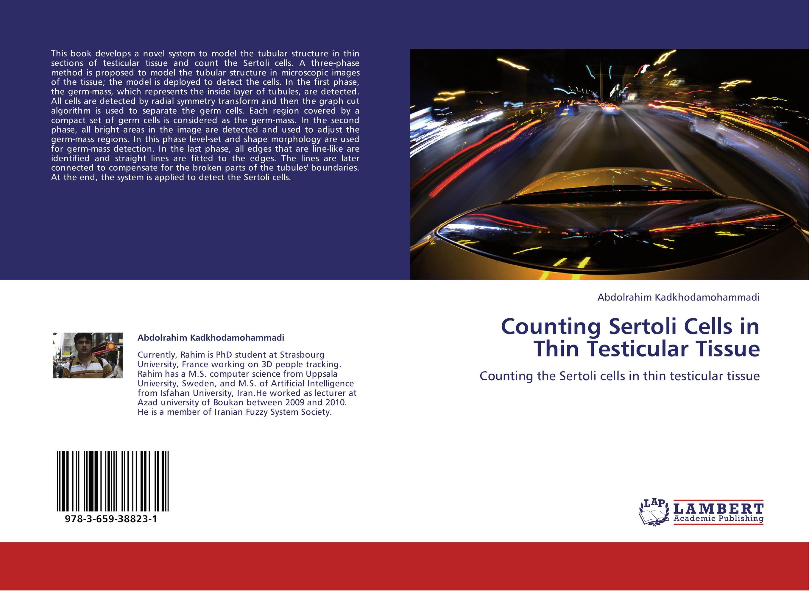
Counting Sertoli Cells in Thin Testicular Tissue. Counting the Sertoli cells in thin testicular tissue

В наличии
| Местонахождение: Алматы | Состояние экземпляра: новый |

Бумажная
версия
версия
Автор: Abdolrahim Kadkhodamohammadi
ISBN: 9783659388231
Год издания: 2013
Формат книги: 60×90/16 (145×215 мм)
Количество страниц: 72
Издательство: LAP LAMBERT Academic Publishing
Цена: 23635 тг
Положить в корзину
| Способы доставки в город Алматы * комплектация (срок до отгрузки) не более 2 рабочих дней |
| Самовывоз из города Алматы (пункты самовывоза партнёра CDEK) |
| Курьерская доставка CDEK из города Москва |
| Доставка Почтой России из города Москва |
Аннотация: This book develops a novel system to model the tubular structure in thin sections of testicular tissue and count the Sertoli cells. A three-phase method is proposed to model the tubular structure in microscopic images of the tissue; the model is deployed to detect the cells. In the first phase, the germ-mass, which represents the inside layer of tubules, are detected. All cells are detected by radial symmetry transform and then the graph cut algorithm is used to separate the germ cells. Each region covered by a compact set of germ cells is considered as the germ-mass. In the second phase, all bright areas in the image are detected and used to adjust the germ-mass regions. In this phase level-set and shape morphology are used for germ-mass detection. In the last phase, all edges that are line-like are identified and straight lines are fitted to the edges. The lines are later connected to compensate for the broken parts of the tubules' boundaries. At the end, the system is applied to detect the Sertoli cells.
Ключевые слова: Cells segmentation, Modelling tubular structure, and Graph cuts.


