| Поиск по каталогу |
|
(строгое соответствие)
|
- Профессиональная
- Научно-популярная
- Художественная
- Публицистика
- Детская
- Искусство
- Хобби, семья, дом
- Спорт
- Путеводители
- Блокноты, тетради, открытки
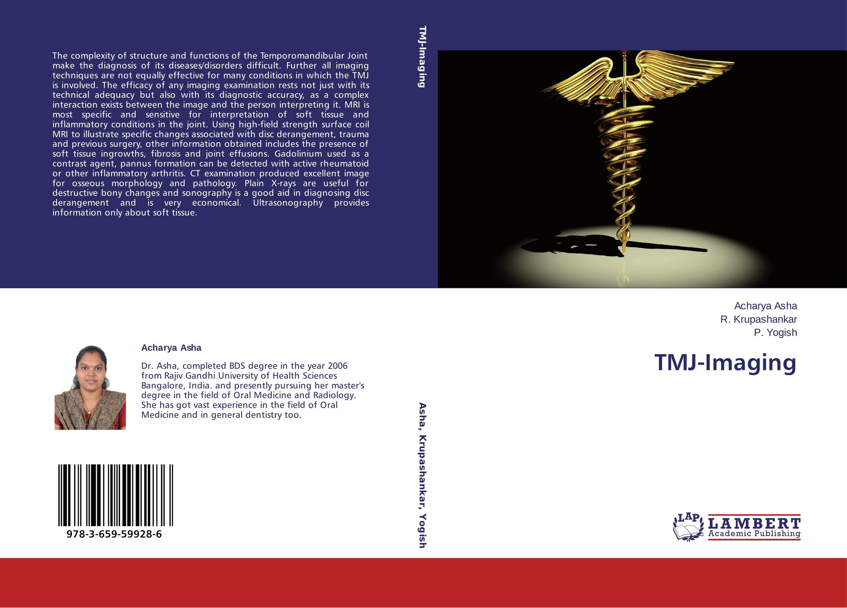
TMJ-Imaging.

В наличии
| Местонахождение: Алматы | Состояние экземпляра: новый |

Бумажная
версия
версия
Автор: Acharya Asha,R. Krupashankar and P. Yogish
ISBN: 9783659599286
Год издания: 2014
Формат книги: 60×90/16 (145×215 мм)
Количество страниц: 176
Издательство: LAP LAMBERT Academic Publishing
Цена: 42817 тг
Положить в корзину
| Способы доставки в город Алматы * комплектация (срок до отгрузки) не более 2 рабочих дней |
| Самовывоз из города Алматы (пункты самовывоза партнёра CDEK) |
| Курьерская доставка CDEK из города Москва |
| Доставка Почтой России из города Москва |
Аннотация: The complexity of structure and functions of the Temporomandibular Joint make the diagnosis of its diseases/disorders difficult. Further all imaging techniques are not equally effective for many conditions in which the TMJ is involved. The efficacy of any imaging examination rests not just with its technical adequacy but also with its diagnostic accuracy, as a complex interaction exists between the image and the person interpreting it. MRI is most specific and sensitive for interpretation of soft tissue and inflammatory conditions in the joint. Using high-field strength surface coil MRI to illustrate specific changes associated with disc derangement, trauma and previous surgery, other information obtained includes the presence of soft tissue ingrowths, fibrosis and joint effusions. Gadolinium used as a contrast agent, pannus formation can be detected with active rheumatoid or other inflammatory arthritis. CT examination produced excellent image for osseous morphology and pathology. Plain X-rays are useful for destructive bony changes and sonography is a good aid in diagnosing disc derangement and is very economical. Ultrasonography provides information only about soft tissue.
Ключевые слова: TMJ imaging


