| Поиск по каталогу |
|
(строгое соответствие)
|
- Профессиональная
- Научно-популярная
- Художественная
- Публицистика
- Детская
- Искусство
- Хобби, семья, дом
- Спорт
- Путеводители
- Блокноты, тетради, открытки
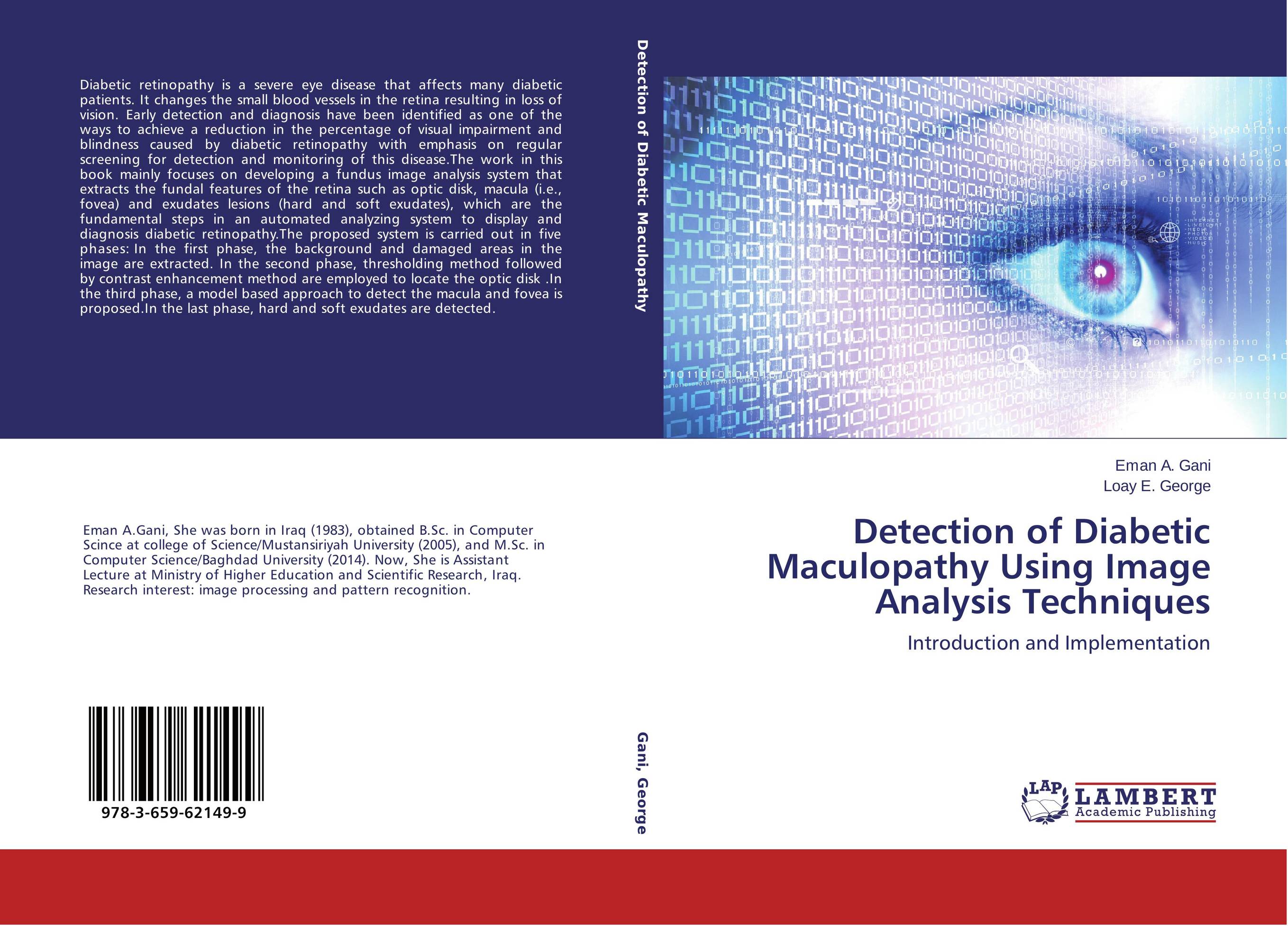
Detection of Diabetic Maculopathy Using Image Analysis Techniques. Introduction and Implementation

В наличии
| Местонахождение: Алматы | Состояние экземпляра: новый |

Бумажная
версия
версия
Автор: Eman A. Gani and Loay E. George
ISBN: 9783659621499
Год издания: 2014
Формат книги: 60×90/16 (145×215 мм)
Количество страниц: 164
Издательство: LAP LAMBERT Academic Publishing
Цена: 42391 тг
Положить в корзину
Позиции в рубрикаторе
Отрасли знаний:Код товара: 141311
| Способы доставки в город Алматы * комплектация (срок до отгрузки) не более 2 рабочих дней |
| Самовывоз из города Алматы (пункты самовывоза партнёра CDEK) |
| Курьерская доставка CDEK из города Москва |
| Доставка Почтой России из города Москва |
Аннотация: Diabetic retinopathy is a severe eye disease that affects many diabetic patients. It changes the small blood vessels in the retina resulting in loss of vision. Early detection and diagnosis have been identified as one of the ways to achieve a reduction in the percentage of visual impairment and blindness caused by diabetic retinopathy with emphasis on regular screening for detection and monitoring of this disease.The work in this book mainly focuses on developing a fundus image analysis system that extracts the fundal features of the retina such as optic disk, macula (i.e., fovea) and exudates lesions (hard and soft exudates), which are the fundamental steps in an automated analyzing system to display and diagnosis diabetic retinopathy.The proposed system is carried out in five phases: In the first phase, the background and damaged areas in the image are extracted. In the second phase, thresholding method followed by contrast enhancement method are employed to locate the optic disk .In the third phase, a model based approach to detect the macula and fovea is proposed.In the last phase, hard and soft exudates are detected.
Ключевые слова: image processing, Image segmentation, Exudates, Diabetic retinopathy, Retinal Image


