


| Поиск по каталогу |
|
(строгое соответствие)
|
- Профессиональная
- Научно-популярная
- Художественная
- Публицистика
- Детская
- Искусство
- Хобби, семья, дом
- Спорт
- Путеводители
- Блокноты, тетради, открытки
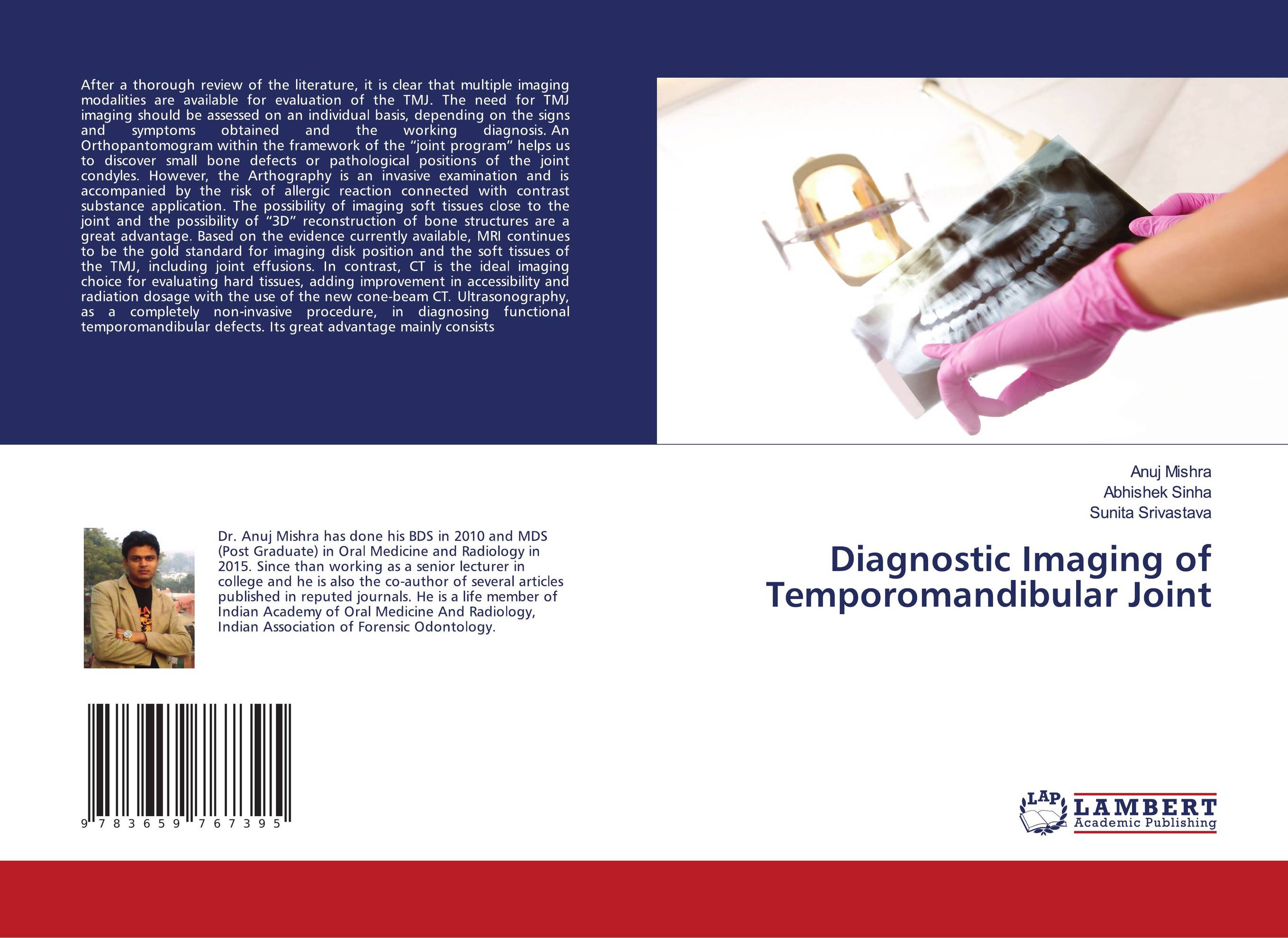
Diagnostic Imaging of Temporomandibular Joint.
В наличии
| Местонахождение: Алматы | Состояние экземпляра: новый |

Бумажная
версия
версия
Автор: Anuj Mishra,Abhishek Sinha and Sunita Srivastava
ISBN: 9783659767395
Год издания: 2015
Формат книги: 60×90/16 (145×215 мм)
Количество страниц: 100
Издательство: LAP LAMBERT Academic Publishing
Цена: 31889 тг
Положить в корзину
| Способы доставки в город Алматы * комплектация (срок до отгрузки) не более 2 рабочих дней |
| Самовывоз из города Алматы (пункты самовывоза партнёра CDEK) |
| Курьерская доставка CDEK из города Москва |
| Доставка Почтой России из города Москва |
Аннотация: After a thorough review of the literature, it is clear that multiple imaging modalities are available for evaluation of the TMJ. The need for TMJ imaging should be assessed on an individual basis, depending on the signs and symptoms obtained and the working diagnosis. An Orthopantomogram within the framework of the “joint program” helps us to discover small bone defects or pathological positions of the joint condyles. However, the Arthography is an invasive examination and is accompanied by the risk of allergic reaction connected with contrast substance application. The possibility of imaging soft tissues close to the joint and the possibility of “3D” reconstruction of bone structures are a great advantage. Based on the evidence currently available, MRI continues to be the gold standard for imaging disk position and the soft tissues of the TMJ, including joint effusions. In contrast, CT is the ideal imaging choice for evaluating hard tissues, adding improvement in accessibility and radiation dosage with the use of the new cone-beam CT. Ultrasonography, as a completely non-invasive procedure, in diagnosing functional temporomandibular defects. Its great advantage mainly consists
Ключевые слова: CBCT, CT, MRI, Temporomandibular joint, Imaging Modalities, TMJ views
Похожие издания
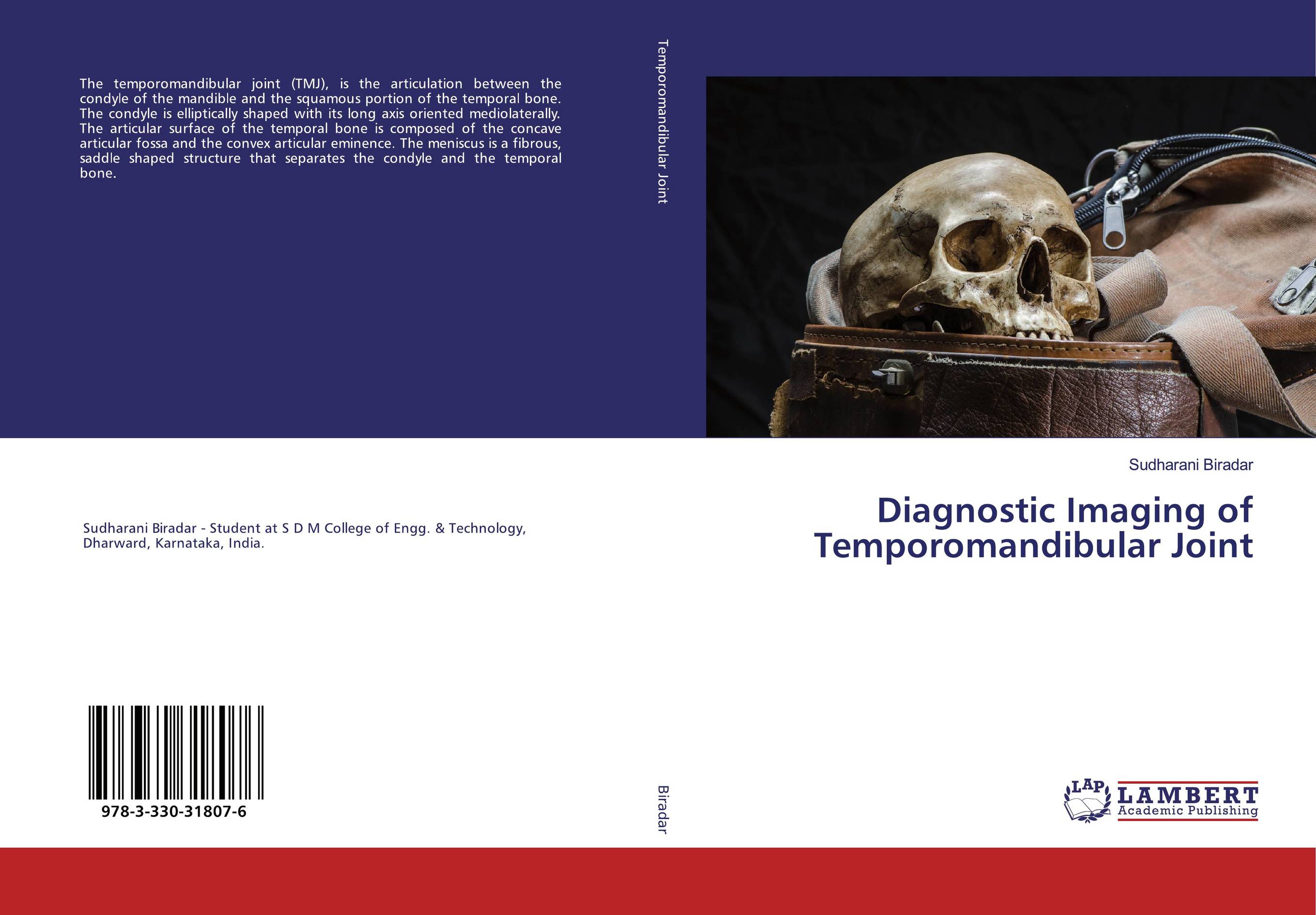 | Отрасли знаний: Общественные науки -> Экономика Sudharani Biradar Diagnostic Imaging of Temporomandibular Joint. . 2019 г., 344 стр., мягкий переплет The temporomandibular joint (TMJ), is the articulation between the condyle of the mandible and the squamous portion of the temporal bone. The condyle is elliptically shaped with its long axis oriented mediolaterally. The articular surface of the temporal bone is composed of the concave articular fossa and the convex articular eminence. The... | 56528 тг |
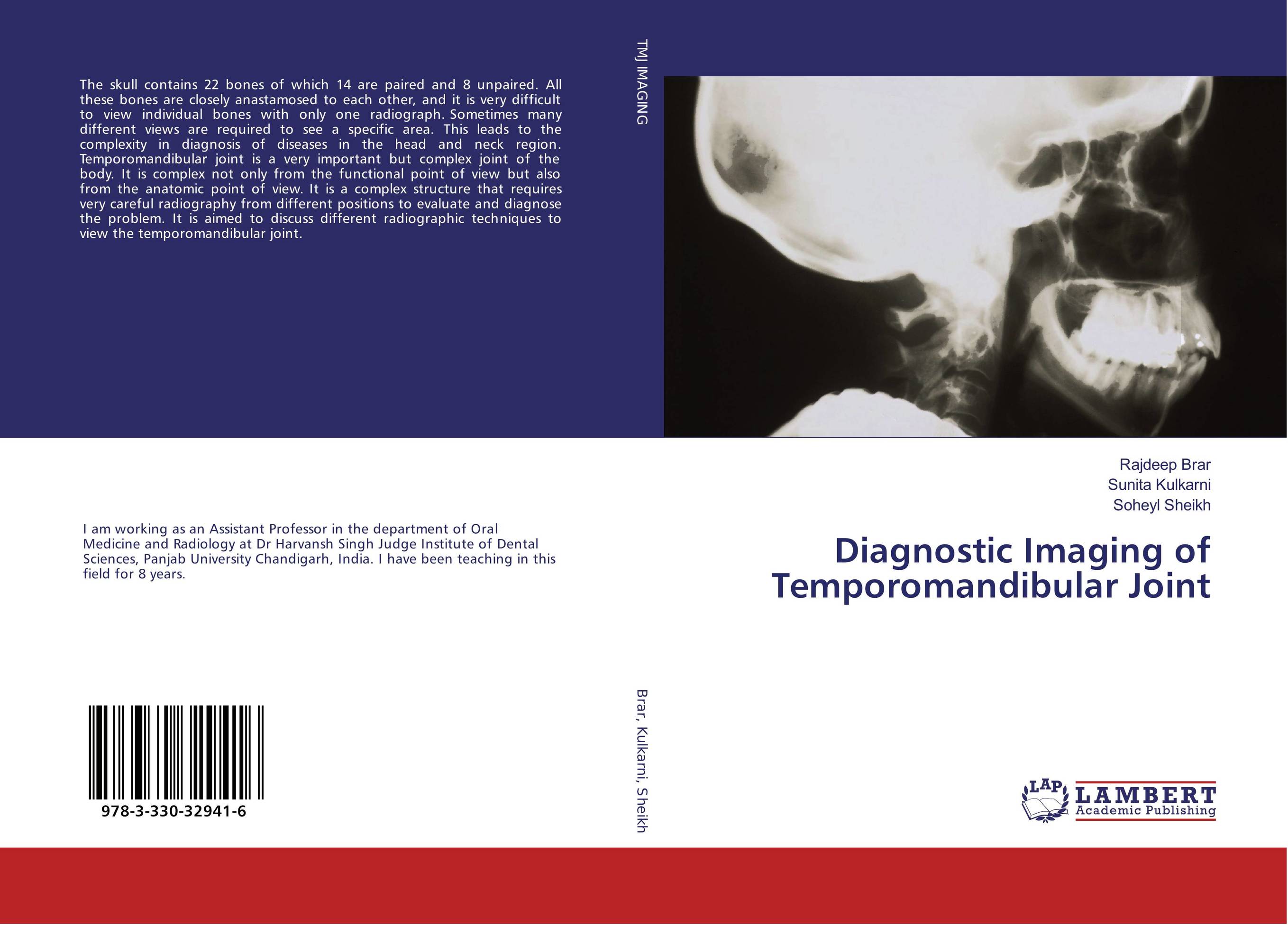 | Отрасли знаний: Медицина -> Стоматология Rajdeep Brar,Sunita Kulkarni and Soheyl Sheikh Diagnostic Imaging of Temporomandibular Joint. . 2017 г., 168 стр., мягкий переплет The skull contains 22 bones of which 14 are paired and 8 unpaired. All these bones are closely anastamosed to each other, and it is very difficult to view individual bones with only one radiograph. Sometimes many different views are required to see a specific area. This leads to the complexity in diagnosis of diseases in the head and neck region.... | 39144 тг |