| Поиск по каталогу |
|
(строгое соответствие)
|
- Профессиональная
- Научно-популярная
- Художественная
- Публицистика
- Детская
- Искусство
- Хобби, семья, дом
- Спорт
- Путеводители
- Блокноты, тетради, открытки
Optimization of double immunofluorescence for cancer tissues.

В наличии
| Местонахождение: Алматы | Состояние экземпляра: новый |

Бумажная
версия
версия
Автор: Natalia Gurvits
ISBN: 9783659621888
Год издания: 2016
Формат книги: 60×90/16 (145×215 мм)
Количество страниц: 88
Издательство: LAP LAMBERT Academic Publishing
Цена: 29043 тг
Положить в корзину
| Способы доставки в город Алматы * комплектация (срок до отгрузки) не более 2 рабочих дней |
| Самовывоз из города Алматы (пункты самовывоза партнёра CDEK) |
| Курьерская доставка CDEK из города Москва |
| Доставка Почтой России из города Москва |
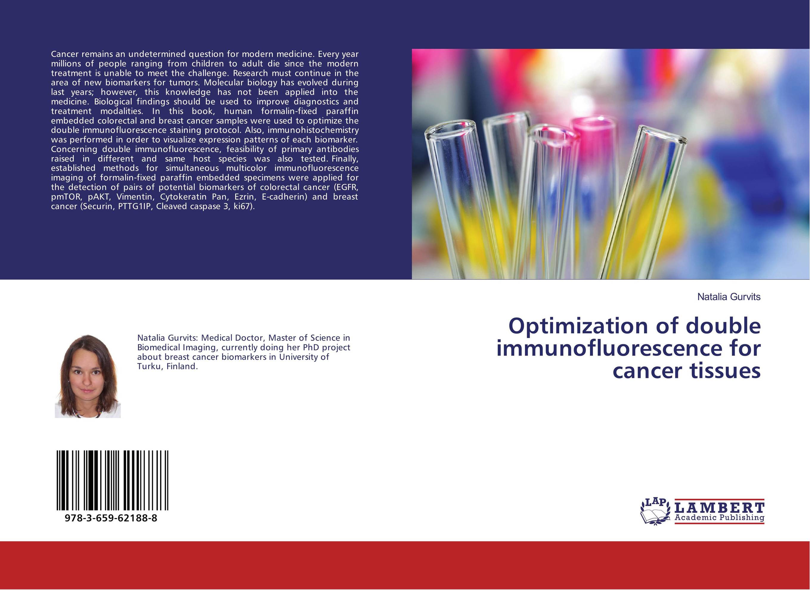
Аннотация: Cancer remains an undetermined question for modern medicine. Every year millions of people ranging from children to adult die since the modern treatment is unable to meet the challenge. Research must continue in the area of new biomarkers for tumors. Molecular biology has evolved during last years; however, this knowledge has not been applied into the medicine. Biological findings should be used to improve diagnostics and treatment modalities. In this book, human formalin-fixed paraffin embedded colorectal and breast cancer samples were used to optimize the double immunofluorescence staining protocol. Also, immunohistochemistry was performed in order to visualize expression patterns of each biomarker. Concerning double immunofluorescence, feasibility of primary antibodies raised in different and same host species was also tested. Finally, established methods for simultaneous multicolor immunofluorescence imaging of formalin-fixed paraffin embedded specimens were applied for the detection of pairs of potential biomarkers of colorectal cancer (EGFR, pmTOR, pAKT, Vimentin, Cytokeratin Pan, Ezrin, E-cadherin) and breast cancer (Securin, PTTG1IP, Cleaved caspase 3, ki67).
Ключевые слова: biomarker, immunohistochemistry, histology, pathology, Cancer, immunofluorescence, microscopy


