| Поиск по каталогу |
|
(строгое соответствие)
|
- Профессиональная
- Научно-популярная
- Художественная
- Публицистика
- Детская
- Искусство
- Хобби, семья, дом
- Спорт
- Путеводители
- Блокноты, тетради, открытки
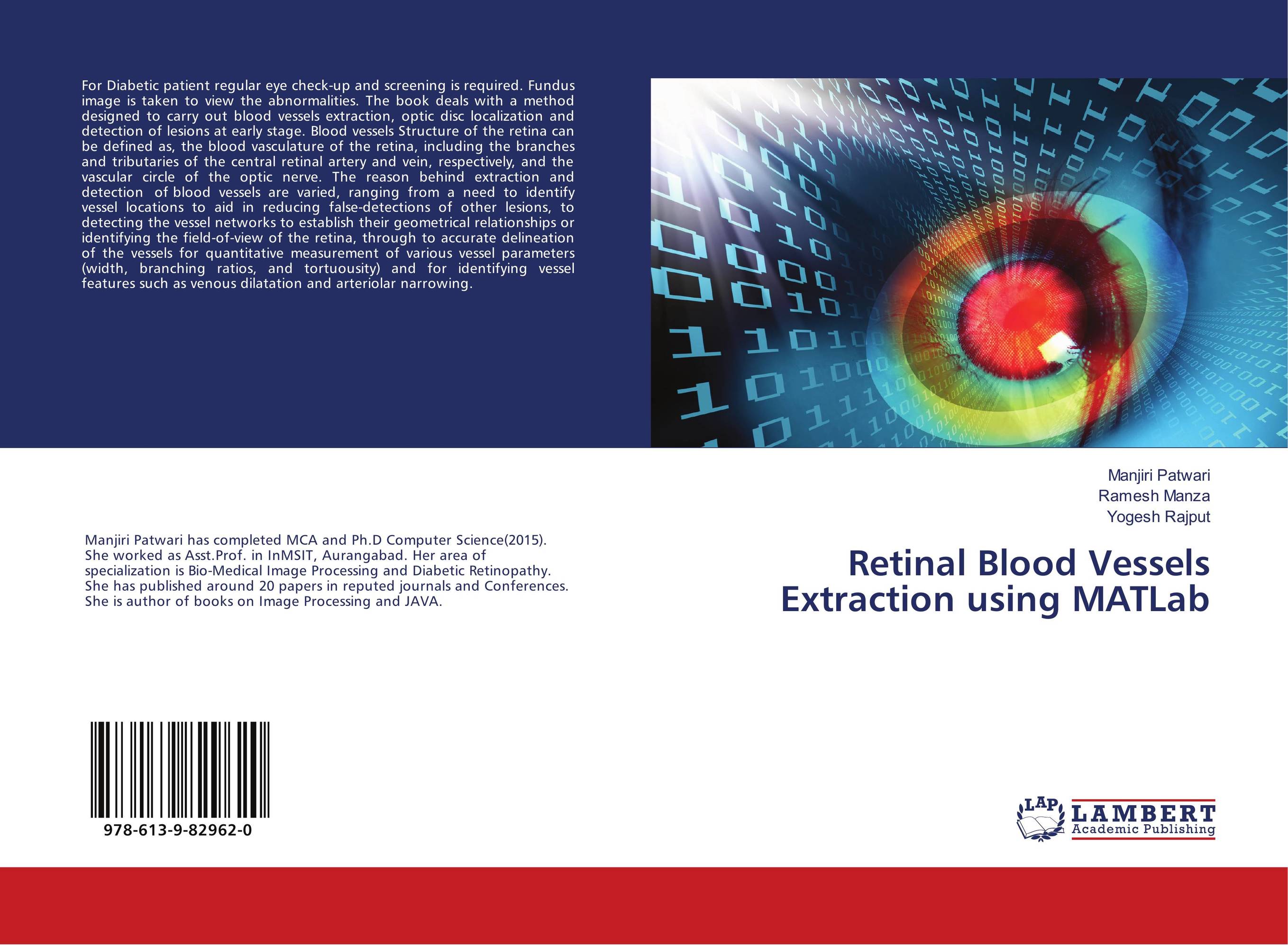
Retinal Blood Vessels Extraction using MATLab.

В наличии
| Местонахождение: Алматы | Состояние экземпляра: новый |

Бумажная
версия
версия
Автор: Manjiri Patwari,Ramesh Manza and Yogesh Rajput
ISBN: 9786139829620
Год издания: 2018
Формат книги: 60×90/16 (145×215 мм)
Количество страниц: 52
Издательство: LAP LAMBERT Academic Publishing
Цена: 20988 тг
Положить в корзину
Позиции в рубрикаторе
Отрасли знаний:Код товара: 206854
| Способы доставки в город Алматы * комплектация (срок до отгрузки) не более 2 рабочих дней |
| Самовывоз из города Алматы (пункты самовывоза партнёра CDEK) |
| Курьерская доставка CDEK из города Москва |
| Доставка Почтой России из города Москва |
Аннотация: For Diabetic patient regular eye check-up and screening is required. Fundus image is taken to view the abnormalities. The book deals with a method designed to carry out blood vessels extraction, optic disc localization and detection of lesions at early stage. Blood vessels Structure of the retina can be defined as, the blood vasculature of the retina, including the branches and tributaries of the central retinal artery and vein, respectively, and the vascular circle of the optic nerve. The reason behind extraction and detection of blood vessels are varied, ranging from a need to identify vessel locations to aid in reducing false-detections of other lesions, to detecting the vessel networks to establish their geometrical relationships or identifying the field-of-view of the retina, through to accurate delineation of the vessels for quantitative measurement of various vessel parameters (width, branching ratios, and tortuousity) and for identifying vessel features such as venous dilatation and arteriolar narrowing.
Ключевые слова: Bio-Medical Image Processing, detection, ?detection, Vessels


