| Поиск по каталогу |
|
(строгое соответствие)
|
- Профессиональная
- Научно-популярная
- Художественная
- Публицистика
- Детская
- Искусство
- Хобби, семья, дом
- Спорт
- Путеводители
- Блокноты, тетради, открытки
Segmentation Based Brain Tumor Detection & Classification.

В наличии
| Местонахождение: Алматы | Состояние экземпляра: новый |

Бумажная
версия
версия
Автор: Anilkumar Suthar and Krunal Patel
ISBN: 9783659514814
Год издания: 2018
Формат книги: 60×90/16 (145×215 мм)
Количество страниц: 56
Издательство: LAP LAMBERT Academic Publishing
Цена: 23066 тг
Положить в корзину
| Способы доставки в город Алматы * комплектация (срок до отгрузки) не более 2 рабочих дней |
| Самовывоз из города Алматы (пункты самовывоза партнёра CDEK) |
| Курьерская доставка CDEK из города Москва |
| Доставка Почтой России из города Москва |
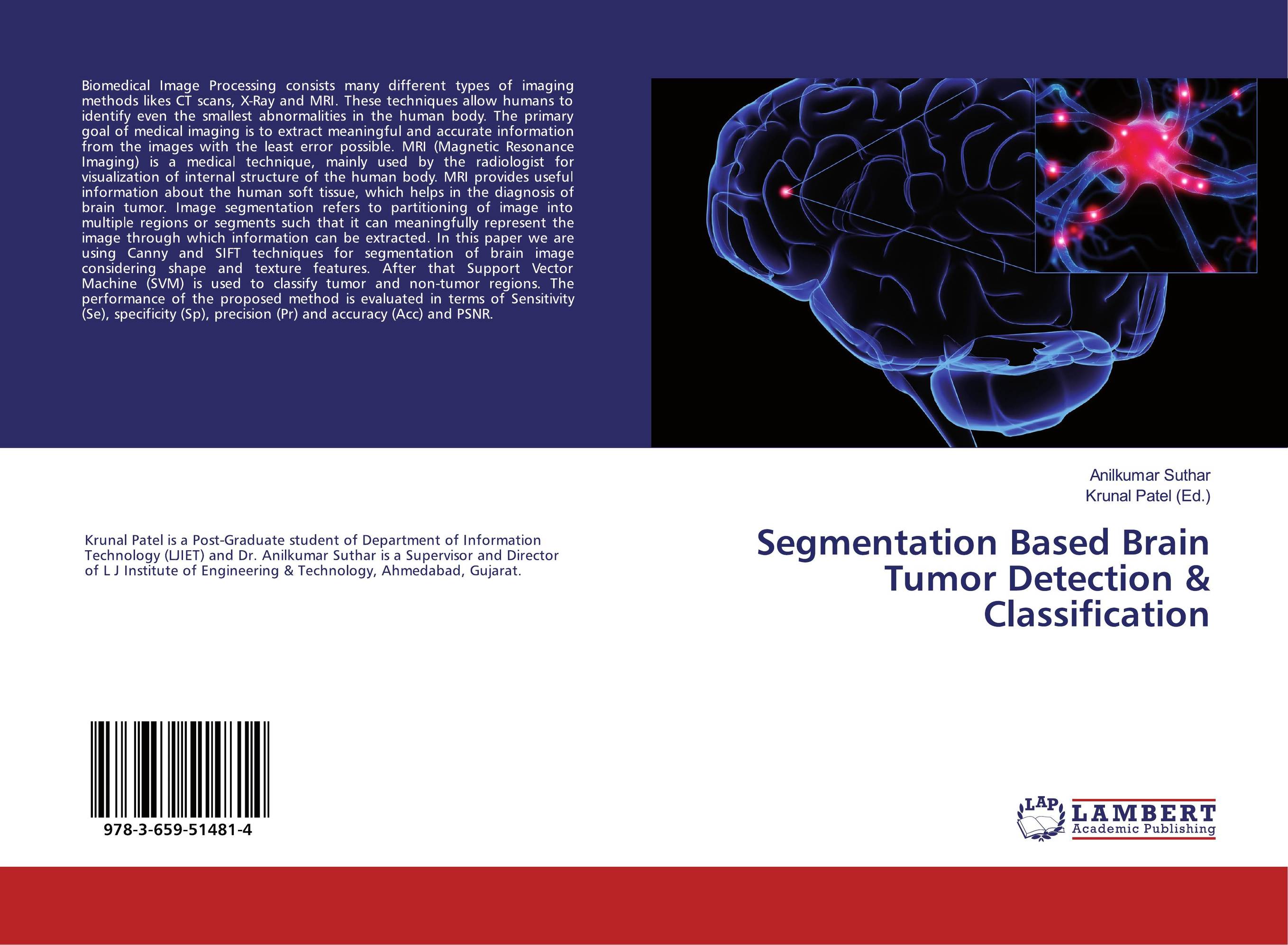
Аннотация: Biomedical Image Processing consists many different types of imaging methods likes CT scans, X-Ray and MRI. These techniques allow humans to identify even the smallest abnormalities in the human body. The primary goal of medical imaging is to extract meaningful and accurate information from the images with the least error possible. MRI (Magnetic Resonance Imaging) is a medical technique, mainly used by the radiologist for visualization of internal structure of the human body. MRI provides useful information about the human soft tissue, which helps in the diagnosis of brain tumor. Image segmentation refers to partitioning of image into multiple regions or segments such that it can meaningfully represent the image through which information can be extracted. In this paper we are using Canny and SIFT techniques for segmentation of brain image considering shape and texture features. After that Support Vector Machine (SVM) is used to classify tumor and non-tumor regions. The performance of the proposed method is evaluated in terms of Sensitivity (Se), specificity (Sp), precision (Pr) and accuracy (Acc) and PSNR.
Ключевые слова: MRI Images, Brain Tumor Detection, segmentation, Brain tumor extraction, classification


