| Поиск по каталогу |
|
(строгое соответствие)
|
- Профессиональная
- Научно-популярная
- Художественная
- Публицистика
- Детская
- Искусство
- Хобби, семья, дом
- Спорт
- Путеводители
- Блокноты, тетради, открытки
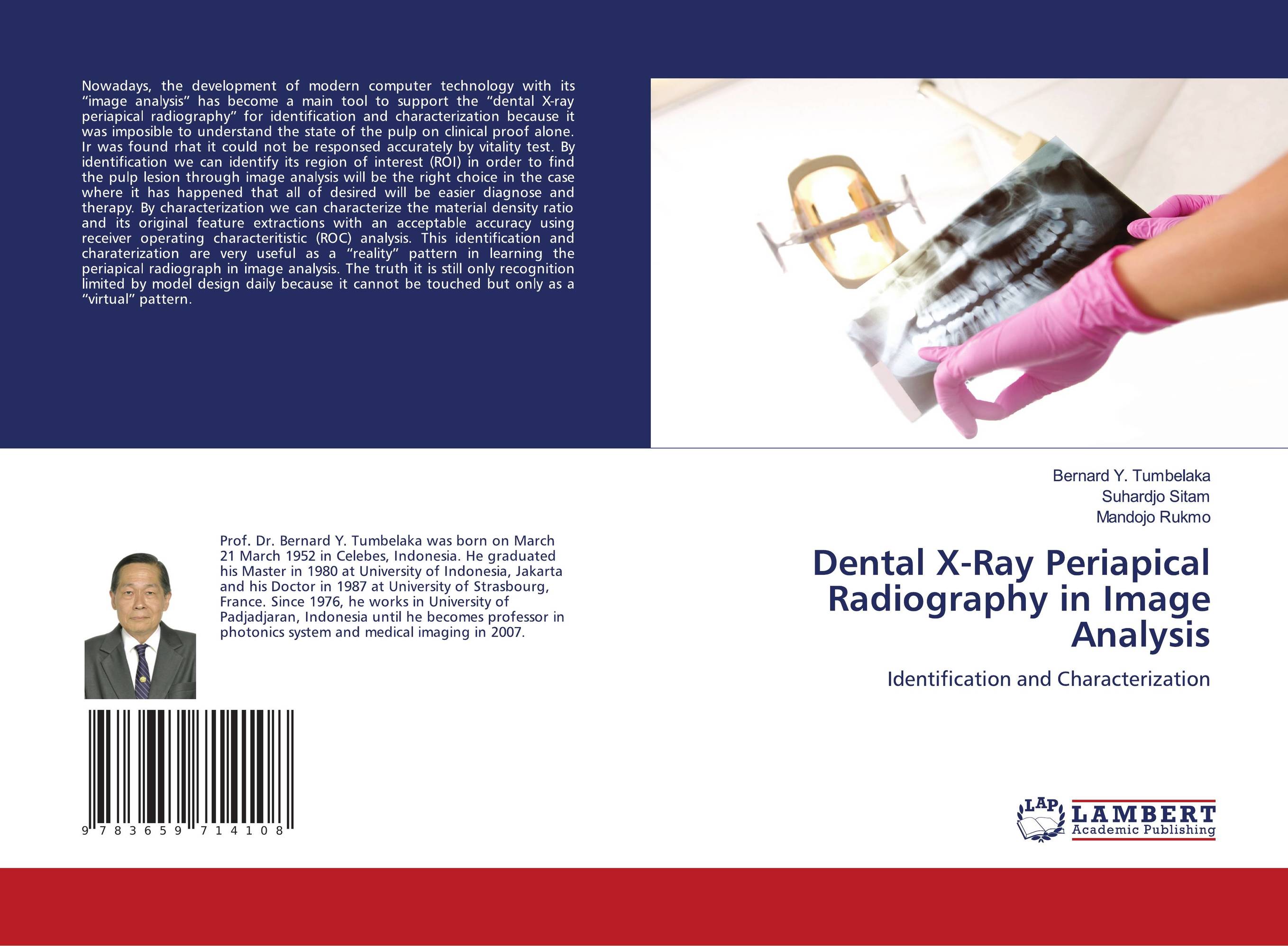
Dental X-Ray Periapical Radiography in Image Anaysis. Identification and Characterization

В наличии
| Местонахождение: Алматы | Состояние экземпляра: новый |

Бумажная
версия
версия
Автор: Bernard Y. Tumbelaka,Suhardjo Sitam and Mandojo Rukmo
ISBN: 9783659714108
Год издания: 2018
Формат книги: 60×90/16 (145×215 мм)
Количество страниц: 52
Издательство: LAP LAMBERT Academic Publishing
Цена: 22924 тг
Положить в корзину
| Способы доставки в город Алматы * комплектация (срок до отгрузки) не более 2 рабочих дней |
| Самовывоз из города Алматы (пункты самовывоза партнёра CDEK) |
| Курьерская доставка CDEK из города Москва |
| Доставка Почтой России из города Москва |
Аннотация: Nowadays, the development of modern computer technology with its “image analysis” has become a main tool to support the “dental X-ray periapical radiography” for identification and characterization because it was imposible to understand the state of the pulp on clinical proof alone. Ir was found rhat it could not be responsed accurately by vitality test. By identification we can identify its region of interest (ROI) in order to find the pulp lesion through image analysis will be the right choice in the case where it has happened that all of desired will be easier diagnose and therapy. By characterization we can characterize the material density ratio and its original feature extractions with an acceptable accuracy using receiver operating characteritistic (ROC) analysis. This identification and charaterization are very useful as a “reality” pattern in learning the periapical radiograph in image analysis. The truth it is still only recognition limited by model design daily because it cannot be touched but only as a “virtual” pattern.
Ключевые слова: identification, charaterization, dental X-ray, periapical radiography


