| Поиск по каталогу |
|
(строгое соответствие)
|
- Профессиональная
- Научно-популярная
- Художественная
- Публицистика
- Детская
- Искусство
- Хобби, семья, дом
- Спорт
- Путеводители
- Блокноты, тетради, открытки
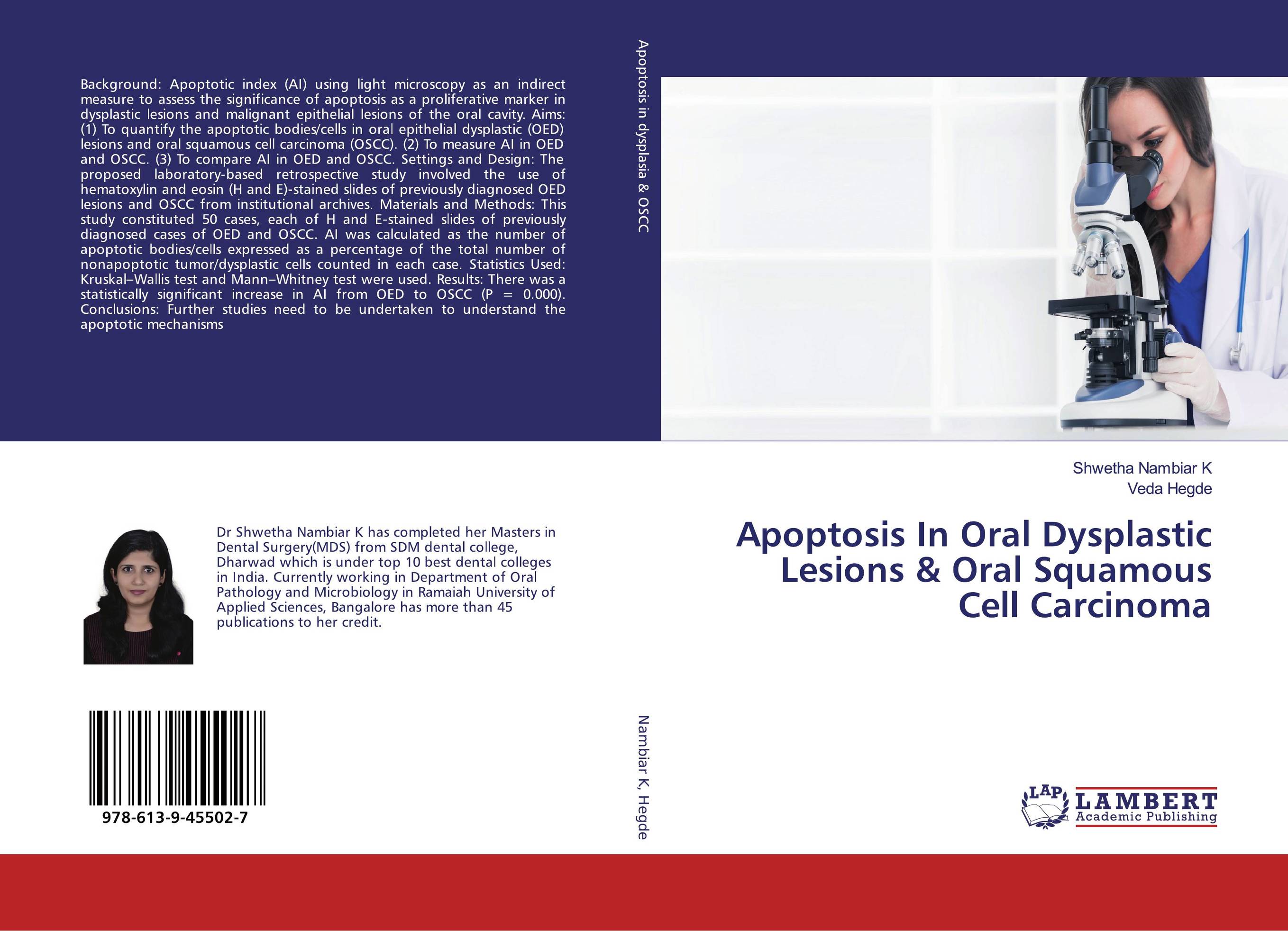
Apoptosis In Oral Dysplastic Lesions & Oral Squamous Cell Carcinoma.

В наличии
| Местонахождение: Алматы | Состояние экземпляра: новый |

Бумажная
версия
версия
Автор: Shwetha Nambiar K and Veda Hegde
ISBN: 9786139455027
Год издания: 2019
Формат книги: 60×90/16 (145×215 мм)
Количество страниц: 136
Издательство: LAP LAMBERT Academic Publishing
Цена: 36556 тг
Положить в корзину
| Способы доставки в город Алматы * комплектация (срок до отгрузки) не более 2 рабочих дней |
| Самовывоз из города Алматы (пункты самовывоза партнёра CDEK) |
| Курьерская доставка CDEK из города Москва |
| Доставка Почтой России из города Москва |
Аннотация: Background: Apoptotic index (AI) using light microscopy as an indirect measure to assess the significance of apoptosis as a proliferative marker in dysplastic lesions and malignant epithelial lesions of the oral cavity. Aims: (1) To quantify the apoptotic bodies/cells in oral epithelial dysplastic (OED) lesions and oral squamous cell carcinoma (OSCC). (2) To measure AI in OED and OSCC. (3) To compare AI in OED and OSCC. Settings and Design: The proposed laboratory?based retrospective study involved the use of hematoxylin and eosin (H and E)?stained slides of previously diagnosed OED lesions and OSCC from institutional archives. Materials and Methods: This study constituted 50 cases, each of H and E?stained slides of previously diagnosed cases of OED and OSCC. AI was calculated as the number of apoptotic bodies/cells expressed as a percentage of the total number of nonapoptotic tumor/dysplastic cells counted in each case. Statistics Used: Kruskal–Wallis test and Mann–Whitney test were used. Results: There was a statistically significant increase in AI from OED to OSCC (P = 0.000). Conclusions: Further studies need to be undertaken to understand the apoptotic mechanisms
Ключевые слова: apoptosis, microscopy, Oral dysplastic lesions, oral squamous cell carcinoma


