


| Поиск по каталогу |
|
(строгое соответствие)
|
- Профессиональная
- Научно-популярная
- Художественная
- Публицистика
- Детская
- Искусство
- Хобби, семья, дом
- Спорт
- Путеводители
- Блокноты, тетради, открытки
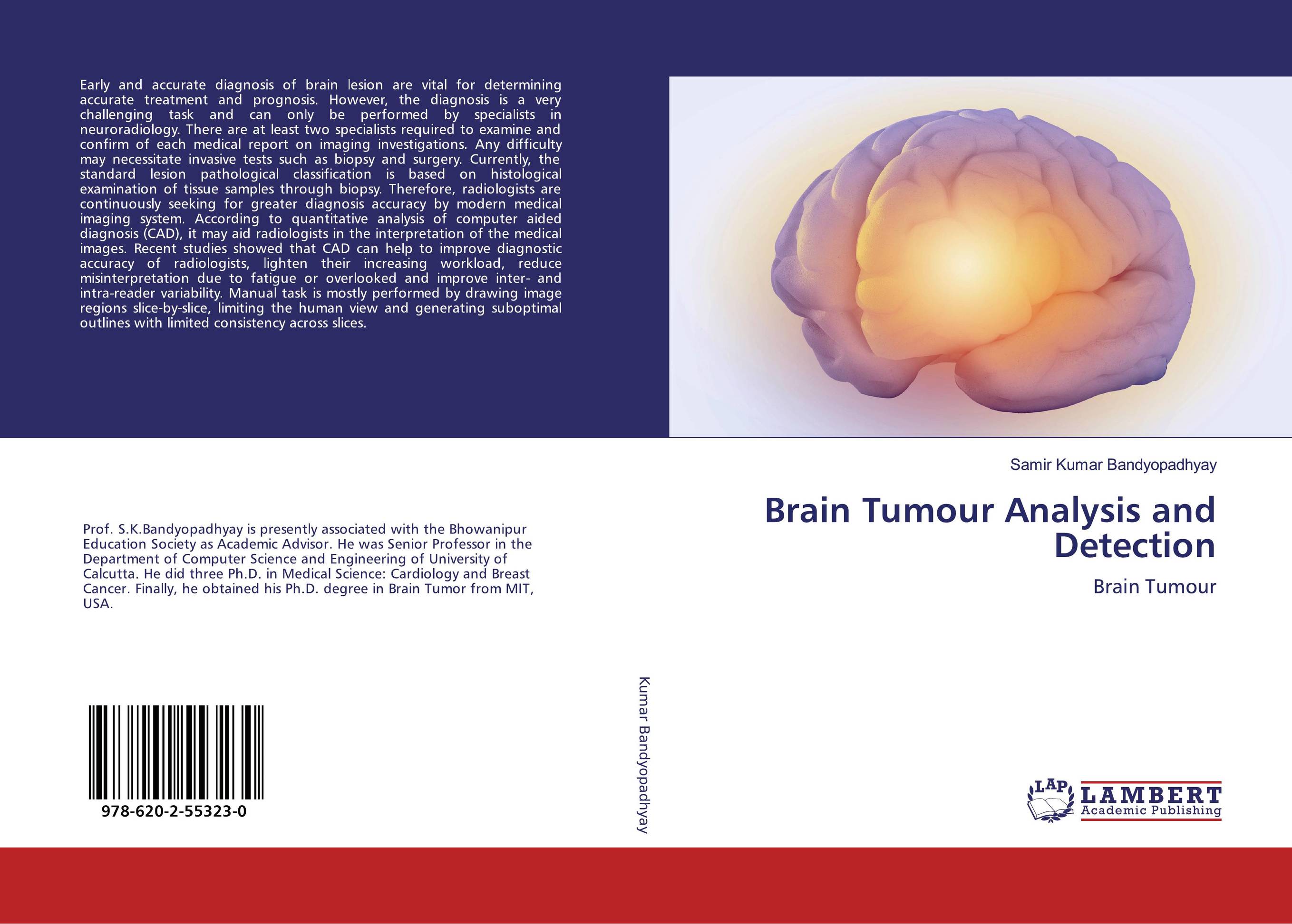
Brain Tumour Analysis and Detection. Brain Tumour
В наличии
| Местонахождение: Алматы | Состояние экземпляра: новый |

Бумажная
версия
версия
Автор: Samir Kumar Bandyopadhyay
ISBN: 9786202553230
Год издания: 2020
Формат книги: 60×90/16 (145×215 мм)
Количество страниц: 188
Издательство: LAP LAMBERT Academic Publishing
Цена: 43243 тг
Положить в корзину
| Способы доставки в город Алматы * комплектация (срок до отгрузки) не более 2 рабочих дней |
| Самовывоз из города Алматы (пункты самовывоза партнёра CDEK) |
| Курьерская доставка CDEK из города Москва |
| Доставка Почтой России из города Москва |
Аннотация: Early and accurate diagnosis of brain lesion are vital for determining accurate treatment and prognosis. However, the diagnosis is a very challenging task and can only be performed by specialists in neuroradiology. There are at least two specialists required to examine and confirm of each medical report on imaging investigations. Any difficulty may necessitate invasive tests such as biopsy and surgery. Currently, the standard lesion pathological classification is based on histological examination of tissue samples through biopsy. Therefore, radiologists are continuously seeking for greater diagnosis accuracy by modern medical imaging system. According to quantitative analysis of computer aided diagnosis (CAD), it may aid radiologists in the interpretation of the medical images. Recent studies showed that CAD can help to improve diagnostic accuracy of radiologists, lighten their increasing workload, reduce misinterpretation due to fatigue or overlooked and improve inter- and intra-reader variability. Manual task is mostly performed by drawing image regions slice-by-slice, limiting the human view and generating suboptimal outlines with limited consistency across slices.
Ключевые слова: BRAIN TUMOUR, Diagnostic CAD, MR images, imaging investigations