| Поиск по каталогу |
|
(строгое соответствие)
|
- Профессиональная
- Научно-популярная
- Художественная
- Публицистика
- Детская
- Искусство
- Хобби, семья, дом
- Спорт
- Путеводители
- Блокноты, тетради, открытки
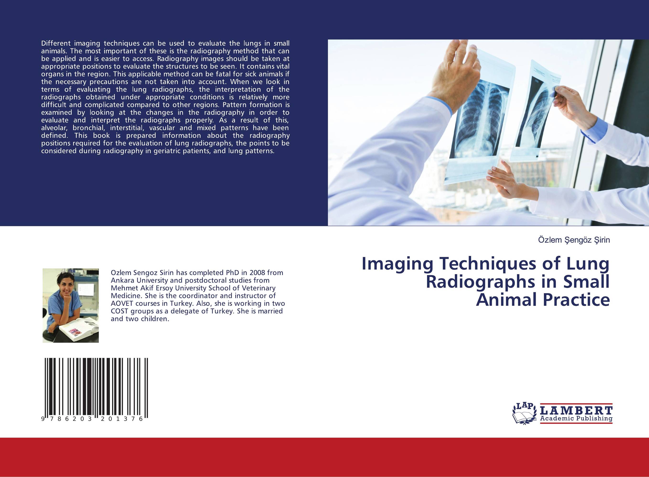
Imaging Techniques of Lung Radiographs in Small Animal Practice.

В наличии
| Местонахождение: Алматы | Состояние экземпляра: новый |

Бумажная
версия
версия
Автор: ?zlem ?eng?z ?irin
ISBN: 9786203201376
Год издания: 1905
Формат книги: 60×90/16 (145×215 мм)
Количество страниц: 52
Издательство: LAP LAMBERT Academic Publishing
Цена: 22924 тг
Положить в корзину
| Способы доставки в город Алматы * комплектация (срок до отгрузки) не более 2 рабочих дней |
| Самовывоз из города Алматы (пункты самовывоза партнёра CDEK) |
| Курьерская доставка CDEK из города Москва |
| Доставка Почтой России из города Москва |
Аннотация: The first radiological examination to be performed in the evaluation of thoracic pathologies is direct radiographs. In order to interpret mediastinal pathologies radiologically, anatomical structures in radiographs should be well recognized. Direct graph is the first method chosen because it is easy to obtain. However, since it displays a volume data on a two-dimensional film plane, the densities of anatomical formations and pathologies are represented on top of each other. This situation is called superposition and constitutes the most important disadvantage of this method. Images should be taken at appropriate positions to evaluate the structures to be seen. This applicable method could be fatal for sick animals if the necessary precautions are not taken into account. Interpretation of the lung radiographs obtained in the correct positions is relatively difficult and complex compared to other regions. This book is prepared to give information about the radiography positions required for the evaluation of lung radiographs, the points to be considered during radiography in esp.geriatric patients, and lung patterns.
Ключевые слова: Lung patterns, Imaging techniques, Small Animal, positions


