| Поиск по каталогу |
|
(строгое соответствие)
|
- Профессиональная
- Научно-популярная
- Художественная
- Публицистика
- Детская
- Искусство
- Хобби, семья, дом
- Спорт
- Путеводители
- Блокноты, тетради, открытки
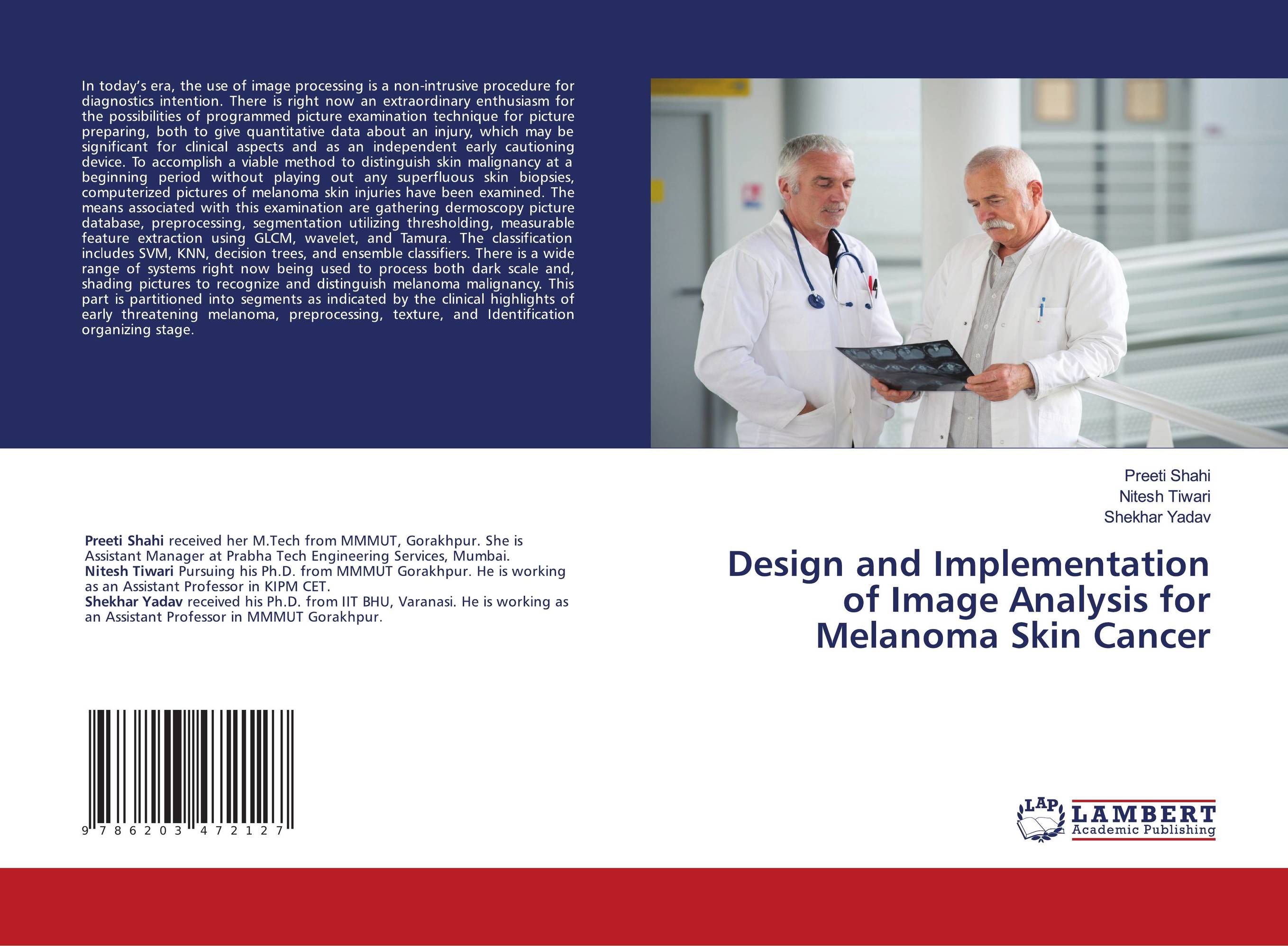
Design and Implementation of Image Analysis for Melanoma Skin Cancer.

В наличии
| Местонахождение: Алматы | Состояние экземпляра: новый |

Бумажная
версия
версия
Автор: Preeti Shahi,Nitesh Tiwari and Shekhar Yadav
ISBN: 9786203472127
Год издания: 1905
Формат книги: 60×90/16 (145×215 мм)
Количество страниц: 52
Издательство: LAP LAMBERT Academic Publishing
Цена: 22924 тг
Положить в корзину
| Способы доставки в город Алматы * комплектация (срок до отгрузки) не более 2 рабочих дней |
| Самовывоз из города Алматы (пункты самовывоза партнёра CDEK) |
| Курьерская доставка CDEK из города Москва |
| Доставка Почтой России из города Москва |
Аннотация: In today’s era, the use of image processing is a non-intrusive procedure for diagnostics intention. There is right now an extraordinary enthusiasm for the possibilities of programmed picture examination technique for picture preparing, both to give quantitative data about an injury, which may be significant for clinical aspects and as an independent early cautioning device. To accomplish a viable method to distinguish skin malignancy at a beginning period without playing out any superfluous skin biopsies, computerized pictures of melanoma skin injuries have been examined. The means associated with this examination are gathering dermoscopy picture database, preprocessing, segmentation utilizing thresholding, measurable feature extraction using GLCM, wavelet, and Tamura. The classification includes SVM, KNN, decision trees, and ensemble classifiers. There is a wide range of systems right now being used to process both dark scale and, shading pictures to recognize and distinguish melanoma malignancy. This part is partitioned into segments as indicated by the clinical highlights of early threatening melanoma, preprocessing, texture, and Identification organizing stage.
Ключевые слова: skin cancer, malignancy, GLCM, SVM, image processing


