| Поиск по каталогу |
|
(строгое соответствие)
|
- Профессиональная
- Научно-популярная
- Художественная
- Публицистика
- Детская
- Искусство
- Хобби, семья, дом
- Спорт
- Путеводители
- Блокноты, тетради, открытки
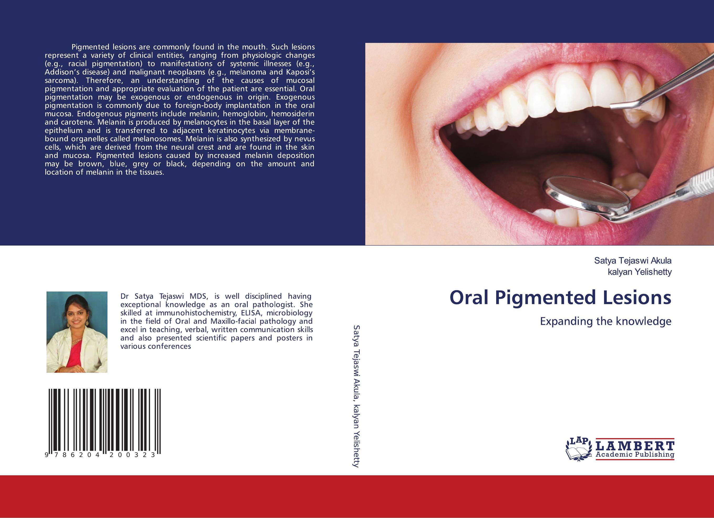
Oral Pigmented Lesions. Expanding the knowledge

В наличии
| Местонахождение: Алматы | Состояние экземпляра: новый |

Бумажная
версия
версия
Автор: Satya Tejaswi Akula and kalyan Yelishetty
ISBN: 9786204200323
Год издания: 1905
Формат книги: 60×90/16 (145×215 мм)
Количество страниц: 120
Издательство: LAP LAMBERT Academic Publishing
Цена: 32599 тг
Положить в корзину
| Способы доставки в город Алматы * комплектация (срок до отгрузки) не более 2 рабочих дней |
| Самовывоз из города Алматы (пункты самовывоза партнёра CDEK) |
| Курьерская доставка CDEK из города Москва |
| Доставка Почтой России из города Москва |
Аннотация: Pigmented lesions are commonly found in the mouth. Such lesions represent a variety of clinical entities, ranging from physiologic changes (e.g., racial pigmentation) to manifestations of systemic illnesses (e.g., Addison’s disease) and malignant neoplasms (e.g., melanoma and Kaposi’s sarcoma). Therefore, an understanding of the causes of mucosal pigmentation and appropriate evaluation of the patient are essential. Oral pigmentation may be exogenous or endogenous in origin. Exogenous pigmentation is commonly due to foreign-body implantation in the oral mucosa. Endogenous pigments include melanin, hemoglobin, hemosiderin and carotene. Melanin is produced by melanocytes in the basal layer of the epithelium and is transferred to adjacent keratinocytes via membrane-bound organelles called melanosomes. Melanin is also synthesized by nevus cells, which are derived from the neural crest and are found in the skin and mucosa. Pigmented lesions caused by increased melanin deposition may be brown, blue, grey or black, depending on the amount and location of melanin in the tissues.
Ключевые слова: Lesions, Oral lesions, oral pigmented lesions
Похожие издания
 | Отрасли знаний: Медицина -> Стоматология Satya Tejaswi Akula and Kalyan Yellisetty Oral Pigmented Lesions.. 1905 г., 148 стр., мягкий переплет The mucous membrane that lines the oral cavity consists of a layer of stratified squamous epithelium and a subepithelial layer, the lamina propria, which consists of a fibrous connective tissue and contain capillaries, nerves and the minor salivary glands. It is not uniformly colored and several degrees of chromatic variation may be observed in... | 36982 тг |


