| Поиск по каталогу |
|
(строгое соответствие)
|
- Профессиональная
- Научно-популярная
- Художественная
- Публицистика
- Детская
- Искусство
- Хобби, семья, дом
- Спорт
- Путеводители
- Блокноты, тетради, открытки
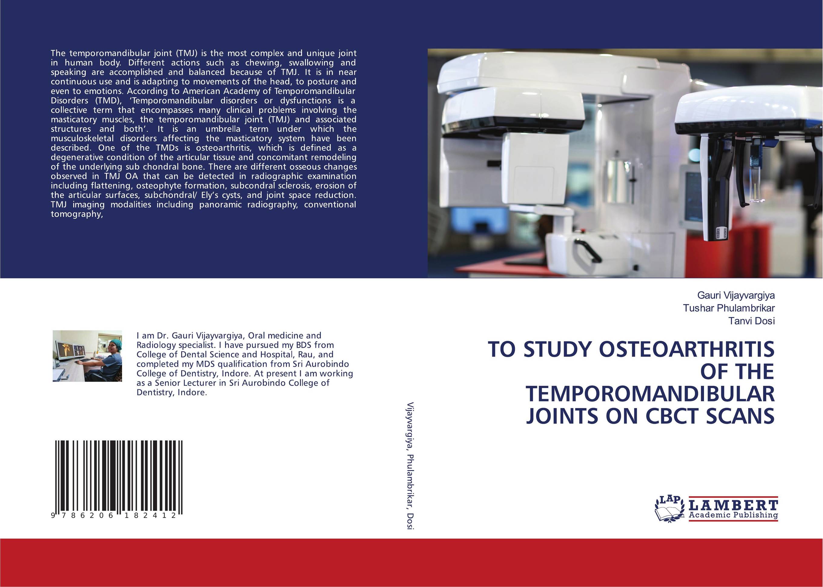
TO STUDY OSTEOARTHRITIS OF THE TEMPOROMANDIBULAR JOINTS ON CBCT SCANS.

В наличии
| Местонахождение: Алматы | Состояние экземпляра: новый |

Бумажная
версия
версия
Автор: Gauri Vijayvargiya,TUSHAR PHULAMBRIKAR and Tanvi Dosi
ISBN: 9786206182412
Год издания: 1905
Формат книги: 60×90/16 (145×215 мм)
Количество страниц: 208
Издательство: LAP LAMBERT Academic Publishing
Цена: 47825 тг
Положить в корзину
| Способы доставки в город Алматы * комплектация (срок до отгрузки) не более 2 рабочих дней |
| Самовывоз из города Алматы (пункты самовывоза партнёра CDEK) |
| Курьерская доставка CDEK из города Москва |
| Доставка Почтой России из города Москва |
Аннотация: The temporomandibular joint (TMJ) is the most complex and unique joint in human body. Different actions such as chewing, swallowing and speaking are accomplished and balanced because of TMJ. It is in near continuous use and is adapting to movements of the head, to posture and even to emotions. According to American Academy of Temporomandibular Disorders (TMD), ‘Temporomandibular disorders or dysfunctions is a collective term that encompasses many clinical problems involving the masticatory muscles, the temporomandibular joint (TMJ) and associated structures and both’. It is an umbrella term under which the musculoskeletal disorders affecting the masticatory system have been described. One of the TMDs is osteoarthritis, which is defined as a degenerative condition of the articular tissue and concomitant remodeling of the underlying sub chondral bone. There are different osseous changes observed in TMJ OA that can be detected in radiographic examination including flattening, osteophyte formation, subcondral sclerosis, erosion of the articular surfaces, subchondral/ Ely’s cysts, and joint space reduction. TMJ imaging modalities including panoramic radiography, conventional tomography,
Ключевые слова: TMJ, osteoarthritis, CBCT


