


| Поиск по каталогу |
|
(строгое соответствие)
|
- Профессиональная
- Научно-популярная
- Художественная
- Публицистика
- Детская
- Искусство
- Хобби, семья, дом
- Спорт
- Путеводители
- Блокноты, тетради, открытки
MRI Image Segmentation for Detection of Brain Tumors.
В наличии
| Местонахождение: Алматы | Состояние экземпляра: новый |

Бумажная
версия
версия
Автор: Chindam Hari Prasad
ISBN: 9786206750918
Год издания: 1905
Формат книги: 60×90/16 (145×215 мм)
Количество страниц: 84
Издательство: LAP LAMBERT Academic Publishing
Цена: 25997 тг
Положить в корзину
Позиции в рубрикаторе
Отрасли знаний:Код товара: 760883
| Способы доставки в город Алматы * комплектация (срок до отгрузки) не более 2 рабочих дней |
| Самовывоз из города Алматы (пункты самовывоза партнёра CDEK) |
| Курьерская доставка CDEK из города Москва |
| Доставка Почтой России из города Москва |
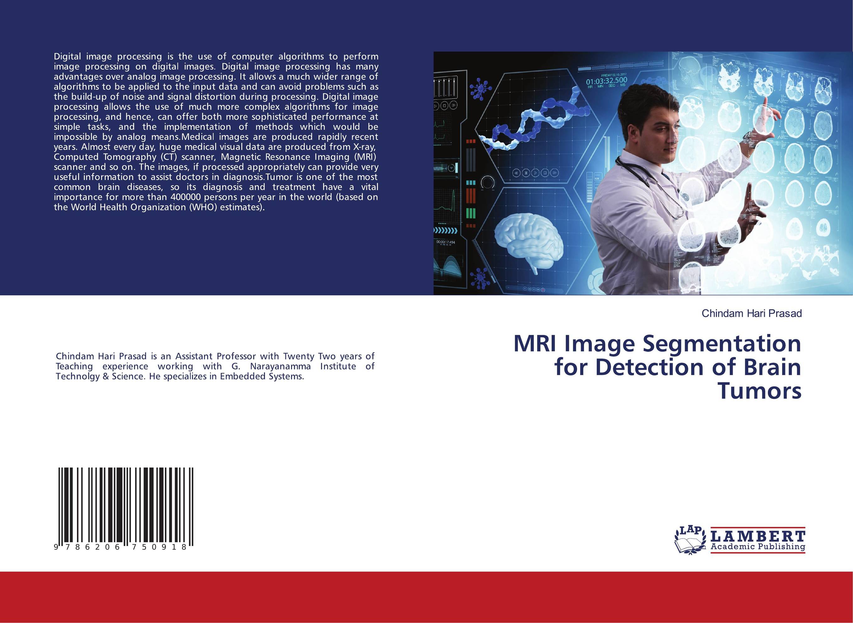
Аннотация: Digital image processing is the use of computer algorithms to perform image processing on digital images. Digital image processing has many advantages over analog image processing. It allows a much wider range of algorithms to be applied to the input data and can avoid problems such as the build-up of noise and signal distortion during processing. Digital image processing allows the use of much more complex algorithms for image processing, and hence, can offer both more sophisticated performance at simple tasks, and the implementation of methods which would be impossible by analog means.Medical images are produced rapidly recent years. Almost every day, huge medical visual data are produced from X-ray, Computed Tomography (CT) scanner, Magnetic Resonance Imaging (MRI) scanner and so on. The images, if processed appropriately can provide very useful information to assist doctors in diagnosis.Tumor is one of the most common brain diseases, so its diagnosis and treatment have a vital importance for more than 400000 persons per year in the world (based on the World Health Organization (WHO) estimates).
Ключевые слова: biomedical, engineering