


| Поиск по каталогу |
|
(строгое соответствие)
|
- Профессиональная
- Научно-популярная
- Художественная
- Публицистика
- Детская
- Искусство
- Хобби, семья, дом
- Спорт
- Путеводители
- Блокноты, тетради, открытки
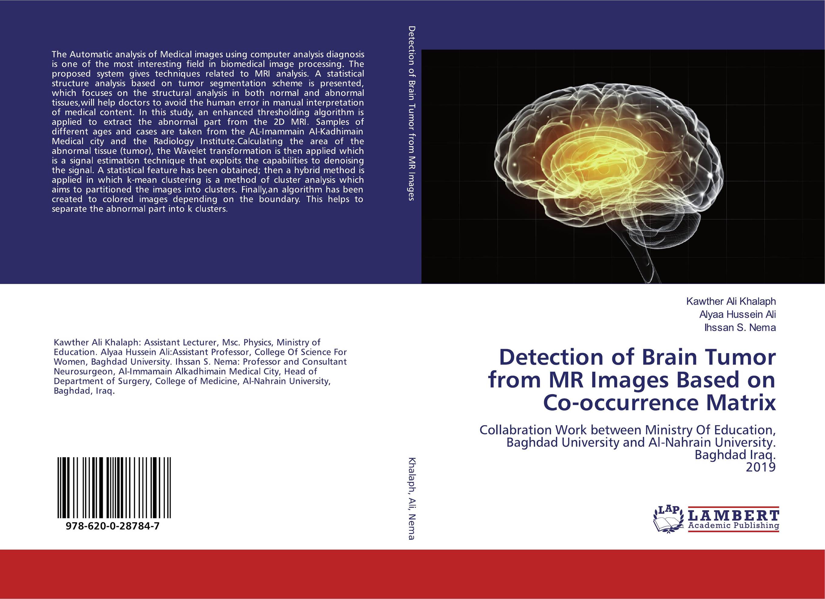
Detection of Brain Tumor from MR Images Based on Co-occurrence Matrix. Collabration Work between Ministry Of Education, Baghdad University and Al-Nahrain University. Baghdad Iraq. 2019
В наличии
| Местонахождение: Алматы | Состояние экземпляра: новый |

Бумажная
версия
версия
Автор: Kawther Ali Khalaph,Alyaa Hussein Ali and Ihssan S. Nema
ISBN: 9786200287847
Год издания: 2019
Формат книги: 60×90/16 (145×215 мм)
Количество страниц: 108
Издательство: LAP LAMBERT Academic Publishing
Цена: 32173 тг
Положить в корзину
| Способы доставки в город Алматы * комплектация (срок до отгрузки) не более 2 рабочих дней |
| Самовывоз из города Алматы (пункты самовывоза партнёра CDEK) |
| Курьерская доставка CDEK из города Москва |
| Доставка Почтой России из города Москва |
Аннотация: The Automatic analysis of Medical images using computer analysis diagnosis is one of the most interesting field in biomedical image processing. The proposed system gives techniques related to MRI analysis. A statistical structure analysis based on tumor segmentation scheme is presented, which focuses on the structural analysis in both normal and abnormal tissues,will help doctors to avoid the human error in manual interpretation of medical content. In this study, an enhanced thresholding algorithm is applied to extract the abnormal part from the 2D MRI. Samples of different ages and cases are taken from the AL-Imammain Al-Kadhimain Medical city and the Radiology Institute.Calculating the area of the abnormal tissue (tumor), the Wavelet transformation is then applied which is a signal estimation technique that exploits the capabilities to denoising the signal. A statistical feature has been obtained; then a hybrid method is applied in which k-mean clustering is a method of cluster analysis which aims to partitioned the images into clusters. Finally,an algorithm has been created to colored images depending on the boundary. This helps to separate the abnormal part into k clusters.
Ключевые слова: Brain Tumor, watershed segmentation, Wavelet Transformation
Похожие издания
 | Отрасли знаний: Точные науки -> Информатика и программирование Chindam Hari Prasad MRI Image Segmentation for Detection of Brain Tumors.. 1905 г., 84 стр., мягкий переплет Digital image processing is the use of computer algorithms to perform image processing on digital images. Digital image processing has many advantages over analog image processing. It allows a much wider range of algorithms to be applied to the input data and can avoid problems such as the build-up of noise and signal distortion during processing.... | 25997 тг |
 | Отрасли экономики: Промышленность в целом Mayur Tiwari Segmentation and Detection of Brain Tumor in Magnetic Resonance Images. Gabor Wavelet Transform Approach. 2017 г., 76 стр., мягкий переплет In medical image processing Segmentation of anatomical regions of brain is the fundamental problem. As the brain structure is very complex involving white matter (WM), gray matter (GM), and cerebrospinal fluid (CSF) this makes feature extraction of brain images as a basic work. Recently MR images are handled manually for the diagnosis of brain... | 21841 тг |